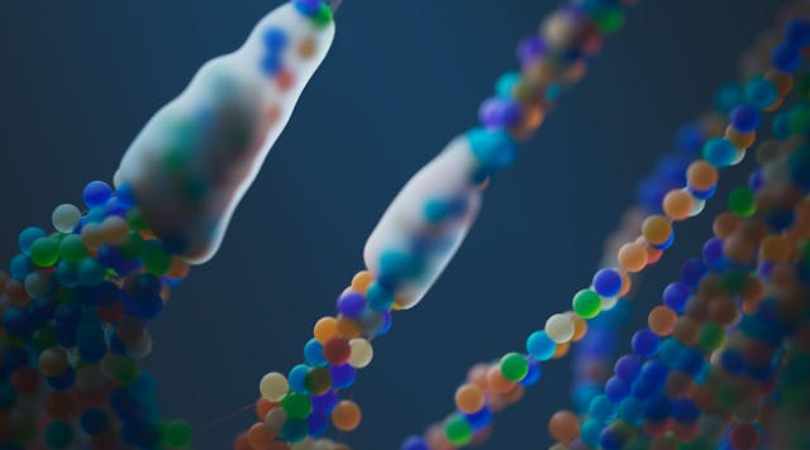
Scientists have successfully restored the lost uricase enzyme, a key breakthrough in combating fructose-induced fat formation. This discovery offers new hope for preventing obesity and metabolic disorders by targeting how the body processes sugar and stores fat.
Limited Quantities Available! Order Today and Enjoy Free Shipping on Orders Over $100!
6.3 Neurodegeneration: The Brain on Fragile Energy
Updated at:

Abstract
The brain is the most energy-demanding organ in the body, consuming about 20% of total resting energy while representing only 2% of body mass. This dependence makes neurons uniquely vulnerable to metabolic disruptions. Dietary fructose itself barely crosses the blood–brain barrier, but the danger arises when neurons generate endogenous fructose internally via the polyol pathway. Triggered by high glucose, salt, dehydration, hypoxia, or stress, this mechanism depletes ATP, raises uric acid, and induces neuronal insulin resistance — a pattern that mirrors the “type 3 diabetes” fingerprint observed in Alzheimer’s disease [NEURO-J2020].
1. The High-Energy Organ at Risk
The brain relies almost entirely on glucose and mitochondrial oxidative phosphorylation for ATP generation. It cannot efficiently burn fat and stores only limited glycogen. Any disturbance in this system — impaired perfusion, mitochondrial dysfunction, or ATP loss — leads to rapid functional decline, manifesting as brain fog, fatigue, or cognitive impairment. The Fructose Model identifies endogenous fructose metabolism within neurons as a critical but overlooked source of this energy instability [NEURO-H2017].
2. Mechanism: Fructose in the Brain
2.1 Endogenous fructose, not dietary fructose
Although dietary fructose does not significantly cross the blood–brain barrier, neurons synthesize fructose locally via the polyol pathway (glucose → sorbitol → fructose). This occurs under stressors such as high glucose spikes, salty carbohydrate-rich meals, dehydration, and hypoxia [ENDO-L2013] [ENDO-AH2021].
2.2 Neuronal insulin resistance
Once activated, fructokinase rapidly consumes ATP, generating uric acid and inducing oxidative stress. This process leads to neuronal insulin resistance — impairing glucose uptake, synaptic plasticity, and memory formation. The result is a brain that remains “hungry” despite energy abundance [NEURO-J2023].
2.3 ATP depletion and mitochondrial suppression
Fructose metabolism causes a sudden ATP drop and mitochondrial downregulation. For neurons, which cannot tolerate energy pauses, this translates into impaired signaling, reduced neurotransmitter turnover, and long-term fragility [MECH-J2007].
2.4 Oxidative stress and blood-flow decline
Uric acid and reactive oxygen species lower nitric oxide, constricting cerebral vessels and compounding energy shortage. Functional imaging confirms that high sugar intake reduces cerebral blood flow in appetite and memory regions [NEURO-P2013].
2.5 Epigenetic instability
Prolonged energy stress disrupts DNA methylation, histone balance, and axonal maintenance. Over time, these molecular alterations accelerate neurodegeneration [NEURO-X2016].
3. From Brain Fog to Alzheimer’s
Animal studies show that high-sugar diets induce neuronal insulin resistance within weeks, progressing to tau tangles and Alzheimer-like pathology by a few months. In human Alzheimer’s brains, researchers have identified elevated fructokinase expression and polyol pathway activation correlating with mitochondrial suppression and cognitive decline [NEURO-J2020]. Epidemiologically, high uric acid levels predict faster cognitive decline and greater dementia risk [NEURO-S2016].
4. Fragile Neurons → Fragile Minds
Energy stress produces diverse outcomes depending on brain region:
- Memory decline: Hippocampal fragility drives Alzheimer’s-like deficits.
- Mood disorders: Low ATP in limbic circuits contributes to anxiety and depression.
- Cognitive fatigue: Energy-starved prefrontal neurons yield brain fog and reduced focus.
- Psychiatric overlap: Emerging reports suggest carbohydrate restriction can restore neuronal stability in select cases of severe psychiatric illness.
Across all, the unifying feature is neuronal energy failure — the same fingerprint as seen in other organs under chronic fructose metabolism.
5. Lessons from Nature: The Arctic Ground Squirrel
During hibernation, Arctic ground squirrels accumulate tau-like proteins similar to those found in Alzheimer’s disease. However, periodic “shivering arousals” restore temperature, energy flow, and protein clearance. When they awaken, their brains are intact. This demonstrates how a temporary, fructose-driven low-energy state can be protective when cyclic — but pathogenic when chronic and unrelieved [NAT-P2017].
6. Beyond Alzheimer’s: A Broader Energy Failure Signature
- Parkinson’s disease: Dopaminergic neurons exhibit mitochondrial fragility and oxidative stress.
- Depression and anxiety: Energy failure disrupts neurotransmitter synthesis and resilience.
- ASD: Altered gut–brain metabolism and mitochondrial stress may involve the same polyol-driven mechanism.
- Schizophrenia: Anecdotal reversals under carbohydrate restriction align with restoration of neuronal energy stability.
Across disorders, the same theme emerges: diverse symptoms, one underlying energy deficit. Fructose metabolism may not cause every case, but it reliably amplifies vulnerability across them [NEURO-J2023].
7. Conclusion
Neurodegeneration is best understood as a failure of energy homeostasis. Fructose metabolism, whether from dietary exposure or endogenous activation, weakens neurons through ATP depletion, insulin resistance, mitochondrial suppression, and reduced blood flow. This lens unites Alzheimer’s, Parkinson’s, mood disorders, and autism under a shared energetic signature.
What begins as adaptive energy conservation becomes destructive when left chronically engaged — a survival program turned against the brain itself.
These relationships form a coherent, testable framework to be addressed in forthcoming experimental protocols.
(Selected sources linked inline; full citations in the Master Bibliography.)
Disclaimer: The information in this blog reflects personal opinions, experiences, and emerging research. It is not intended as medical or professional advice and should not replace consultation with qualified professionals. The accuracy of this content is not guaranteed. Always seek guidance from a licensed expert before making any health-related decisions.


